News

Novel Procedure For Implanting Bio-Loggers In Atlantic Bluefin Tuna
Measuring heart rate using bio-loggers in free swimming fishes to detect their response to environmental impacts is a promising tool in aquaculture. It is of great value to have an insight into what drives migration in fish and the impact this could have on commercial utilization and conservation. Due to the logistic challenges that are involved, especially in the study of physiology of large pelagic fish, it has been vital to design simple, less stressful procedures. To perform an invasive surgery into the body cavity to place a bio-logger close to the heart is a difficult option on a commercial vessel with the risk of harming these large fish.
Measuring heart rate, activity and temperature at different intervals
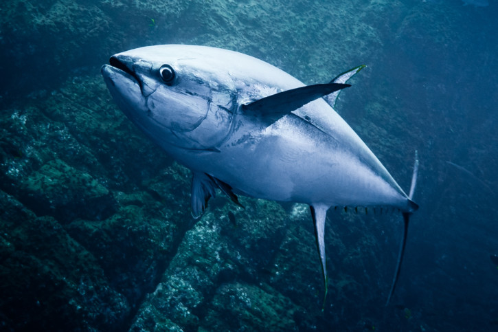
Scientists from France, Denmark, Iceland and Malta implanted Atlantic bluefin tuna (Thunnus thynnus) with Star-Oddi DST centi-HRT ACT with a novel technique, where they used human laparoscopic atraumatic trocar to assist in the implanting process. The study was divided into three different experiments while designing and testing the implanting procedure. The study consisted of three experiments. One short experiment where the tuna was killed after 5 minutes. The second was over 50 days, and in the third the loggers measured for 80 days heart rate, activity and temperature every 10 minutes. In experiment one and two the loggers measured heart rate every 2 minutes.
The protocol for implanting bio-loggers using a laparoscopic trocar
The procedure was performed on conscious fish with covered eyes and sea water flowing over the gills. Each fish was no longer than 2 minutes out of water. The placement of the loggers was aimed in the fleshy area below the operculum in the muscles associated with the cleithrum, which is in close proximity of the heart. First a small incision was made through the skin and then sterilized trocar was inserted into the incision and pushed forward parallel to the lower edge of the operculum, following a natural groove. The depth was carefully assessed depending on the size of the fish, not to damage any vital blood vessels or the heart. No suturing was needed and the healing in experiment 3 was very good. This technique was shown to be promising and easy to repeat and can give good quality data due to its proximity to the heart.

Further results can be found in the article published in Journal of Fish Biology and can be accessed here
